Acetone and cotton swab has been the traditional method for detecting dyed coral. This technique is unfortunately somewhat destructive, removing some of the color from the surface of dyed samples. Raman/PL spectroscopy offers fast non-destructive method for detecting naturally colored coral.
Raman/PL spectra of red coral typically exhibit a mixture of coral matrix (calcium carbonate) and organic unmethylated polyenes (polyacetylenes) or methylated polyenes (carotenoids) responsible for the color. Calcium carbonate may exist in the form of calcite or aragonite. Both naturally colored and dyed corals may exhibit Raman features of these minerals.
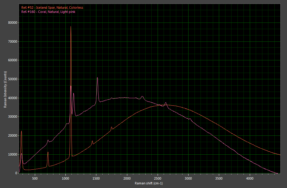
Figure 1: Raman/PL spectra of calcite (Iceland spar) and pink coral showing the peaks of both calcite and polyene compounds.

Naturally colored red coral can be easily identified by the presence of peaks associated to organic pigments. Most important peaks are 1520 cm-1 & 1132 cm-1 and smaller ones at 1297 cm-1 & 1020 cm-1. Broad scan photoluminescence spectrum reveals series of additional Raman peaks caused by the same pigments. Overall appearance of spectrum is characteristic for coral and can only be mistaken for spectra of pink to red pearls (for example conch pearl) . Intensity of the peaks is directly correlated to color saturation of the sample. Dyed coral does not show these peaks although sometimes tiny peaks at differing locations caused by dye substance can be observed riding on the top of broader photoluminescence band.